
What is magnetic resonance imaging?
Magnetic resonance imaging (MRIMRI, or magnetic resonance imaging, is a method of taking 3D images using magnetic fields.) is a procedure that utilizes a strong magnetic field and time-varying electromagnetic waves to obtain images of a patient’s internal anatomy.


Images courtesy of Siemens Healthcare GmbH .
The human body is largely made of water (up to 60%), and each water molecule contains two hydrogen atoms. A hydrogen atom contains a single proton that acts like a compass needle when the atom is in a magnetic field. Normally, the hydrogen protons in our bodies are randomly arranged. When a person’s body is placed in an MRIMRI, or magnetic resonance imaging, is a method of taking 3D images using magnetic fields. scanner, however, the protons align along the direction of the scanner’s strong magnetic field. Then, radiofrequency wavesRadiofrequency waves are electromagnetic wave frequencies that fall in the spectrum of radio transmissions (30 Hz to 300 GHz). Clinical MRIs use frequencies in this range (similar to AM and FM radio), so the electromagnetic wave used in MRI is common... are turned on, making some of the protons change direction. After the radiofrequency wavesRadiofrequency waves are electromagnetic wave frequencies that fall in the spectrum of radio transmissions (30 Hz to 300 GHz). Clinical MRIs use frequencies in this range (similar to AM and FM radio), so the electromagnetic wave used in MRI is common... are switched off, these protons realign with the magnetic field. As the protons realign, they emit their own radio signals which can be detected by an antenna. The intensity and duration of these signals provide information about the location and makeup of tissues in the body, and this information is used to generate 3D images of the patient.
Examples of clinical MRI images.

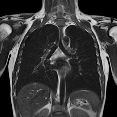

Images courtesy of Siemens Healthcare GmbH.
Different sequences of radiofrequency pulses can be used to highlight different types of tissues, such as muscle, fat, or blood. A wide assortment of pulse sequences and sophisticated signal processing algorithms have been developed to obtain the best possible images of the region of interest in the body. MRIMRI, or magnetic resonance imaging, is a method of taking 3D images using magnetic fields. is a useful tool because it provides high-quality images of soft tissues which are hard to see using other methods, such as x-rays and CT scans. MRIMRI, or magnetic resonance imaging, is a method of taking 3D images using magnetic fields. also does not deliver any radiation dose to the patient.
Why is safety important with MRI?
Because the magnetic field in an MRIMRI, or magnetic resonance imaging, is a method of taking 3D images using magnetic fields. scanner is very strong and always on, certain types of metal items can present a hazard. Objects containing even tiny amounts of ferromagnetic material (such as iron and nickel) can be pulled into the MRIMRI, or magnetic resonance imaging, is a method of taking 3D images using magnetic fields. and injure a patient or worker or damage the machine. Pacemakers, metal clips, and metal valves can potentially be dangerous around MRIMRI, or magnetic resonance imaging, is a method of taking 3D images using magnetic fields. scanners. However, in some cases, patients with these devices can be scanned safely by making small changes to how the scan is performed. It is important for patients to tell the MRIMRI, or magnetic resonance imaging, is a method of taking 3D images using magnetic fields. department about any metal implants they have to make sure the scan can be performed safely.